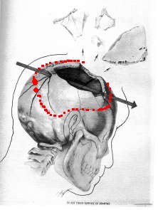
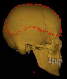
"House Select Committee Medical illustrator Ida Dox, working for the House Select Committee's Forensic Pathology Panel in the late 1970s, produced the drawing [above]. It faithfully reflects what the autopsy photos and x-rays show. " J McAdams:Kennedy Assassination Home Page
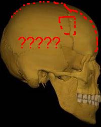
I have taken the liberty of tracing approximately, in red, the outline of the large defect, as described by the Clark Panel and one of the HSCA consultants. The other HSCA consultant seems to have produced a wound compatible with the Dox drawing, but he has added something extra.
He describes a wide crack extending along the centre line of the head right down the forehead and into the right frontal sinus. This is completely contrary to the photographic evidence and witness testimony.
Whether the shot that killed JFK was from the front or the rear it is apparent that the skull beneath the 'intact' scalp on the well known back 'back of the head' photos is, according to these consultants, severely fragmented, displaced outwards , or just plain missing.
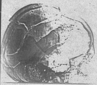
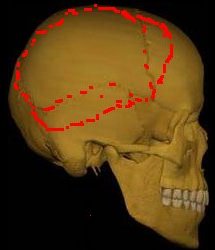
A version of the above drawing that actually shows the damage shown on the xrays is here:

One perfectly viable interpretation of the apparent large pieces of shattered skull on the rear of the Lateral is that, as the autopsy doctors described, the head was falling to pieces in their hands as they worked on it. Between the taking of the lateral (presumably first) and the taking of the AP, these loose pieces had simply fallen off.
The Clark Panel
("1968 Panel Review of Photographs, X-Ray Films, Documents and Other Evidence Pertaining to the Fatal Wounding of President John E Kennedy on November 22, 1963, in Dallas, Texas")
Morgan, Russell H., MD, Professor of Radiology, School of Medicine and Professor of Radiological Sciences, School of Hygiene and Public Health, The Johns Hopkins University, Baltimore, MD,( nominated by Dr. Lincoln Gordon, President of The Johns Hopkins University) concludes that the XRays show: (quote)
"With respect to the right frontoparietal region of the skull, the traumatic damage is particularly severe with extensive fragmentation of the bony structures from the midline of the frontal bone anteriorly to the vicinity of the posterior margin of the parietal bone behind. Above the fragmentation extends approximately 25 mm. across the midline to involve adjacent portions of the left parietal bone; below, the changes extend into the right temporal bone. Throughout this region, many of the bony pieces have bean displaced outward; several pieces are missing." (unquote)
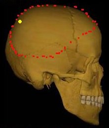
So Russell Morgan puts the 'extensive fragmentation' of the skull back as far as the border of the occipital bone. Note that he is speaking of 'fragmentation' here, not mere fractures. According to my dictionary, the meaning of 'fragmented' is " 1. reduced to fragments. 2. existing or functioning as though broken into separate parts" The picture (right) is based solely on Morgan's description of this 'fragmented' area. Note that we are searching for the solid, intact borders of the large head defect. Clearly in Morgan's opinion the bone is shattered even in the area where his own panel decided there was an entry wound. Note that it is perfectly possible that the back of the head was in fragments and yet be held together by the scalp. (The yellow dot shows the Clark/HSCA 'entry').
The HSCA : 1
Ten years later the consulting radiologist with the HSCA, Dr. G.M. McDonnel, gets to see the XRays: (quote)
"On 7 March 1978, at The Aerospace Corporation, I was asked to interpret six radiographs which are identified by the Number 21296, dated 11/22/63, and Bethesda NMC: "These radiographs were unenhanced. My preliminary interpretation follows: 1. A nearly complete loss of structure of the right frontal and parietal bone." (unquote)
McDonnel is equally explicit. He includes however the whole right frontal in his catalogue of destruction. His description is summarised, right. It is broadly in agreement with Morgan's analysis, and bears not much relation to the Dox drawing.
TheHSCA : 2
By August 4th 1978, McDonnel has seen the enhanced XRays (which he requested), and consulted with colleagues....(quote)
HSCA Exhibit F-33
Report of G.M. McDonnel, M.D. concerning observation, analysis, and conclusions in connection with radiographic images and enhanced images attributed to President John F. Kennedy.
This report replaces my report of March 8, 1978 and supplements my presentation of July 21, 1978 in the Rayburn Building, Washington, D.C.
I was exposed to radiographic images identified by the number 21296 at Aerospace Corporation, El Segundo, California on March 7, 1978. At my suggestion portions of these radiographs were digitized and enhanced by Aerospace Corporation for further observation and analysis.
The findings and interpretation of the skull films are:
1. Nearly complete loss of right parietal bone, the upper portion of the right temporal bone, and a portion of the posterior aspect of the right frontal bone.(unquote)
The area defined by McDonnel is shown at the right.
The HSCA : 3
David O Davis, MD (Professor and Chairman Department of Radiology The George Washington University Hospital Washington DC) also examined the Xrays for the HSCA. His findings on the exact location of the defect are curiously vague. However, Davis adds one particularly startling conclusion: (quote)
August 23, 1978
I reviewed the Kennedy skull films labeled #1 and #2, taken at the US Naval Hospital on September 22, 1963,[sic] and two aerospace enhanced images of those films. The findings are as follows: There is massive calvarial damage, which will be described below.
There is a large fracture extending directly anteriorly along the sagittal suture, which is, at least at the point visualized, widely separated. This fracture seems to extend into the frontal bone, more or less at the midline, down to the frontal sinus which is also fractured. (unquote)
This is quite amazing.
He sees the forehead split in two by this unheretofore mentioned fracture. A glance at one of the autopsy photos will show this to be nonsense.
His wound description is much more vague. I couldn't reproduce it's lower borders accurately from this new description, as he doesn't specify them. (See right) (quote on): CONCLUSION: There is an extensive comminuted, open, explosive calvarial fracture which seems to radiate in various directions as described above from a central point which is located in the right parietal bone, 3cm from the midline and about 9 or 10cm from the external occipital protuberance. There is absence of a part of the calvarium, beginning near the impact point and extending anteriorly to the coronal suture, with absence of a significant amount of bone in the right parietal and presumably a small amount of left parietal region. There is a displaced fragment or fragments in the right frontal and parietotemporal region, with some overlap of the bone.
There is a significant fracture in the frontal region extended into the right orbit and frontal sinus.
The fractures also extend, from the posterior impact point, into the occipital bone on both sides. I neglected to describe in the text of this report an extensive fracture which extends inferolaterally from the impact point toward the left side which probably reaches the temporal bone or at least the mastoid region after crossing a goodly portion of the occipital bone there. It seems apparent that explosive impact occurred in this calvarium. It also seems reasonable to assume that the exit point is near the coronal suture on the right side, about 5 or 6, or perhaps slightly more, cm above the pterion. It is not possible to totally explain the metallic fragment pattern that is present from some of the metallic fragments located superiorly in the region of the parietal bone, or at least projecting on the parietal bone, are actually in the scalp. The frontal view does not give much help in this regard and it is impossible to work this out completely. (unquote)
Note that he dwells at length on the fractures, but is strangely obscure on the 'large defect'. The odd squarish shape in my diagram is a (doomed) attempt to convey this obscurity. The language too is much more hesitant than McDonnels: "probably" "it seems apparent" "it seems reasonable to assume" "it is not possible to explain" "not much help" "can't work this out completely"
Did Davis feel the need for some explanation of the huge chunk of skull apparently missing in the right eye area of the AP? "There is a large fracture extending directly anteriorly along the sagittal suture, which is, at least at the point visualized, widely separated. This fracture seems to extend into the frontal bone, more or less at the midline, down to the frontal sinus which is also fractured."
It seems maybe he did.