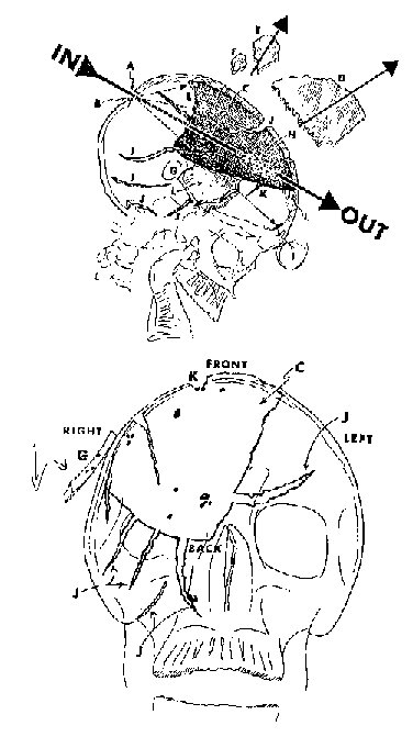
These (above) are diagrams drawn by John Lattimer, a urologist who got to see the x-rays at the archives.
It's obvious from the diagram that Lattimer has the fractures at 'J' at the side of the head (in the lateral) and at the back (in the AP.)
Also check his point labelled 'K'. I have no idea how he came to the conclusion these K's were the same point.
Also, and this is the main point, clearly Lattimer doesn't go along with Joe Durnavich's notion (mine too by this time) that the obvious defect in the AP is mostly just missing brain. He clearly interprets it as missing bone.
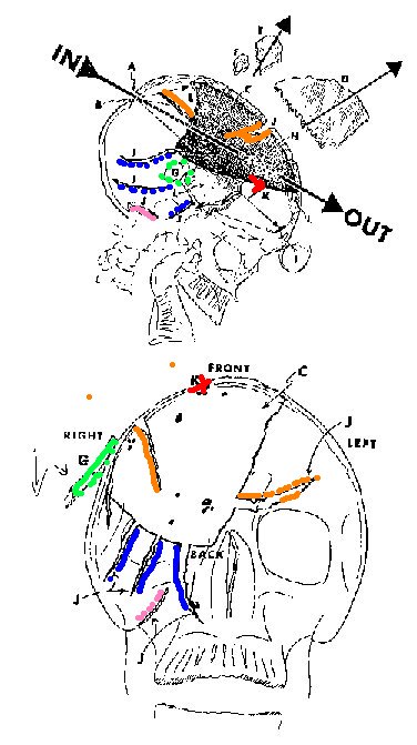 |
Ok. Here's the "Annotated-Colour-Coded-Lattimer-JPG".In both of his pictures he has cracks marked 'J' leading up to the edge of the large defect. With a bit of work, you can match them up as I have done on the left.
If Lattimer didn't intend the lower diagram to show where the missing bone was, how come such a nice correlation between the two? Why did he label all these points with all these J's and Ks if he didn't intend anyone to think he meant them for the same points? |