The Headwound As Seen At Parkland Hospital
"There's no hole in the back of the head there; is there?" (John Stringer, ARRB Deposition)
"Horne stated that you could not tell in the "bootleg" version (the ones we typically see), as it's very dark on the bottom, but the actual image is much better and shows the entire back of the head is lacking in structure. It kind of sags there, as though it kind of caved in where it sits in the stirrup. "
For a more complete & detailed listing compiled by Dr. Gary Aguilar follow this link
Some photographs here have been stolen from Robert Groden's compendious photographic compilation in "The Killing of a President"
The purpose of this page is to illustrate the (apparent) incompatibility between the 'back of the head'
autopsy photographs (below) and the witness testimony. The autopsy photo shows the obvious and universally admitted
'right front' flap. The evidence is that there were in fact three
'major' flaps, one of them being at the right rear.
Dr Boswell - one of the autopsy doctors - maintains that he was lifting such a flap over the back of the head in the photo below.
|
JAMES J. HUMES, MD:
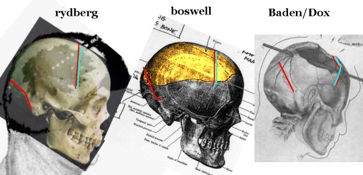
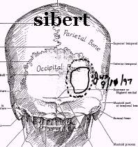
..the other is essentially the leftmost of the three below, though I have superimposed an actual skull on it to make comparisons easier. The defect runs from the borders of the occipital bone into the frontal bone. Boswell produced a drawing for the ARRB (centre, below, defect highlighted) which is very similar. Compare both of these to the Dox drawing produced under Michael Baden's supervision for the HSCA (right,below) which shows the right rear parietal essentially intact. (Solid, though fractured). If the headwound was in fact as in the Rydberg & Boswell drawings, then there is no great puzzle about how the Parkland witnesses could report a 'right rear' defect. The simple fact is, acording to all the evidence, there was one. If the 'Baden/Dox' version is correct , then the radiology is wrong & the Clark Panel were clearly wrong to declare that: " [Autopsy] Photographs 7, 14, 42, and 43 show the back of the head, the contours of which have been grossly distorted by extensive fragmentation of the underlying calvarium." The preceding sentence incidentally makes doubly plain the fact that they believed the 'back of the head' skull was indeed 'fragmented'. " This wound measured approximately 13 centimeters in greatest diameter. It was difficult to measure accurately because radiating at various points from the large defect were multiple crisscrossing fractures of the skull which extended in several directions. I have noted in my report that a detailed description of the lines of these fractures and of the types of fragments that were thus made were very difficult of verbal description, and it was precisely for this reason that the photographs were made so one might appreciate more clearly how much damage had been done to the skull. " (Humes, WC) One searches the Baden/Dox version in vain for any sign of these 'multiple criss-crossing fractures extending in several directions'. See the lateral skull x-ray.
Rydberg drawing 1964 (above, left, skull superimposed) Boswell's drawing for the ARRB. 1998 (above, centre, side view) The HSCA Dox drawing 1978 (right, above) In all 3 pictures, the lamboid suture is highlighted red, and the coronal suture in blue & red.
Humes' comments on autopsy picture # 42 |
||||
|
J. THORNTON BOSWELL, MD:
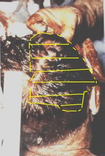
Boswell's drawing for the ARRB (back view, defect highlighted)
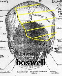
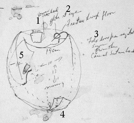
One of Boswell's autopsy diagrams. Head seen from top. Eyes at the top of the diagram. Note the legend: "10 X 17 [cm] missing" in the top of the skull. Boswell maintains that the scalp in the colour autopsy picture (below right) is being pulled forwards over (essentially) a vacancy at the right rear. The brain is removed by the time the picture (below) is taken. This would have have been virtually impossible - & utterly inept - had the back of the head been essentially 'intact' as in the Dox drawing. " It would be irresponsible and stupid to try to remove the brain if so much skull were left, as it must be in the official interpretations of the photographs " according to neuroanatomist Joe Riley (see his article : "What Struck John") Boswell's comments on the autopsy picture
Clearly Dr Boswell is not describing anything like the Baden/Dox version.
|
||||
|
PIERRE A. FINCK, MD:
Like many people, Finck also appears to be having some difficulty with autopsy # 42.
|
||||
| GODFREY McHUGH: President Kennedy's Air Force Aid, described the head wound to David Lifton (BE:430): "...he
was in absolute perfect shape, except the back of the head, top back of the head, had an explosive bullet in it
(sic) and was badly damaged..." To clarify the point Lifton asked: "When you think of the head wounds, then, you think of, primarily, the top of the head, or primarily the back of the head? McHugh answered, "Both. Ninety-nine percent the back, the top back of the head... that's the portion that had been badly damaged by the bullet." (BE:432) Lifton, to leave no doubt about what was meant then asked McHugh to define the back of the head. McHugh answered: "The portion that is in the back of the head, when you're lying down in the bathtub, you hit the back of the head." (Best Evidence, p. 430) Again, this appears to contadict the photo. |
||||
|
JOHN STRINGER: was the autopsy photographer. David Lifton interviewed Stringer, in part, as follows: Lifton: "When you lifted him out, was the main damage to the skull on the top or in the back?" Stringer: "In the back." Lifton: "In the back?...High in the back or lower in the back?" Stringer: "In the occipital part, in the back there, up above the neck." Lifton: "In other words, the main part of his head that was blasted away was in the occipital part of the skull?" Stringer: "Yes. In the back part." Lifton: "The back portion. Okay. In other words, there was no five-inch hole in the top of the skull?" Stringer: "Oh, some of it was blown off--yes, I mean, toward, out of the top in the back, yes." Lifton: "Top in the back. But the top in the front was pretty intact?" Stringer: "Yes, sure." Lifton: "The top front was intact?" Stringer: "Right." Lifton, unsatisfied with precisely what Stringer may have meant by the 'back of the head' asked, as he had asked McHugh, if by "back of the head" Stringer meant the portion of the head that rests on the rear portion of a bathtub during bathing. Stringer replied, "Yes."--as had McHugh (BE, p.516) Stringer On Autopsy Photo #42"There's no hole in the back of the head there; is there?" (John Stringer, ARRB Deposition)
|
||||
| SECRET SERVICE AGENT WILLIAM GREER: was asked by Arlen Specter for the Warren Commission to describe the
head wound he saw at Bethesda. Greer said, "I would--to the best of my recollection it was in this part of
the head right here." Specter immediately asked, "Upper right?" Greer: "Upper right side."
Specter: "Upper right side, going toward the rear. and what was the condition of the skull at that point?"
Greer: "The skull was completely--this part was completely gone." (Warren Comm-- V2:127) |
||||
|
SECRET SERVICE AGENT ROY KELLERMAN: under oath before the Warren Commission explained the head wound he saw to Arlen Specter, "He had a large wound this size." Specter: "Indicating a circle with your finger of the diameter of 5 inches would that be approximately correct?" (sic) Kellerman: "Yes, circular; yes, on this part of the head." Specter: "Indicating the rear portion of the head." Kellerman: "Yes." Specter: "More to the right side of the head." Kellerman: "Right. This was removed." Specter: "When you say, "This was removed", what do you mean by this?" Kellerman: "The skull part was removed." Specter: "All right." Kellerman: "To the left of the (right) ear, sir, and a little high; yes...(I recall that this portion of the rear portion of the skull) was absent when I saw him." (WC-V2:80- 81)
Drawing by Kellerman for the HSCA
|
||||
|
SECRET SERVICE AGENT CLINTON J. HILL: after seeing the President's skull wound in Dealey Plaza, and after returning with the body to Bethesda he was "summoned...down to the morgue to view the body (again) and to witness the damage of the gunshot wounds."--as agent Kellerman put it in his 11-29-63 report. (WC--CE #1024, Kellerman report of 11-29-63. In: WC--V18:26-27) Hill reported, "When I arrived the autopsy had been completed and...I observed another wound (in addition to the throat wound) on the right rear portion of the skull." (WC--CE#1024, V18:744)
|
||||
|
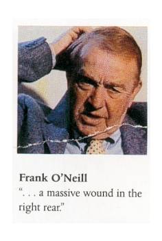
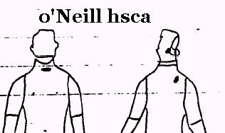
FBI AGENT FRANCIS X. O'NEILL: In an HSCA interview with Andy Purdy and Mark Flanagan on 1/10/78 O'Neill said that the autopsy doctors felt that "the bullet that entered the head struck the center, low portion of the head and exited from the top, right side, towards the front." (HSCA rec # 006185.) However, O'Neill made a sketch witnessed and signed by D. A. Purdy and M. T. Flanagan that showed an "entry" at the low rear central portion of JFK's skull and an 'exit' on the right rear quadrant of the head no more anterior than the posterior portion of the ear. (HSCA rec # 006185 p. 10.) (O'Neill felt it odd that while he had been interviewed by the Warren Commission's Arlen Specter, he had never been called to testify. O'Neill recalled that "On the issue of the full vs. partial autopsy, O'Neill said that Admiral Galloway resolved this by ordering a complete autopsy." (HSCA rec # 006185, p. 3) "O'Neill emphatically stated that the doctors removed only two fragments and not 'a missile'." (IBID. p. 5.) "O'Neill mentioned that the doctors just wanted to obtain the large fragments and that many small fragments did exit.)
O'Neill's drawing for the HSCA
|
||||
|
FBI agent JAMES SIBERT: assisted Francis O'Neill. After an interview for the HSCA J. Kelly and A. Purdy reported, "Regarding the head wound, Sibert said it was in the "...Upper back of the head." (sic) In an affidavit prepared for the HSCA Sibert claimed, "The head wound was in the upper back of the head.", and "...a large head wound in the upper back of the head with a section of the scull (sic) bone missing..." Sibert sketched a drawing of the skull wound and traced a small wound square in the central rear portion of the skull neither to the right or the left, slightly above the level depicted for the ears but well below the level depicted for the top of the skull. (HSCA REC # 002191) (Emphasis added.)
|
||||
|
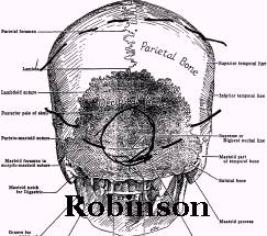
TOM ROBINSON: was the assistant to Joe Hagen, president of Gawler's Funeral Home, which prepared John Kennedy's body for his coffin. Robinson assisted with the preparations for an open casket funeral so preparation of the skull was especially meticulous. Robertson described the skull wound in a 1/12/77 HSCA interview released in 1993 conducted by Andy Purdy and Jim Conzelman: Purdy asked Robinson: "Approximately where was this wound (the skull wound) located?" Robinson: "Directly behind the back of his head." Purdy: "Approximately between the ears or higher up?" Robinson, "No, I would say pretty much between them." (HSCA rec # 189-10089-10178, agency file # 000661, p.3. On the day of their interview Purdy and Conzelman signed a diagram prepared and also signed by Robinson. The sketch depicts a defect directly in the central, lower rear portion of the skull. (HSCA doc # 180-10089-10179, agency file # 000662)
Note that Boswell's autopsy notes also describe the (supposed) eop 'entry' , down near the rear hairline, as 'ragged'.
|
||||
|
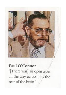
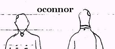
PAUL KELLY O'CONNOR: one of two laboratory technologists present during JFK's autopsy at Bethesda, he has repeatedly insisted that the skull wound extended on the right side well into the rear of the skull. "O'Connor was shown the autopsy photographs and he said, "No, that doesn't look like what I saw...A lot worse wound extended way back here, " and he demonstrated with his hand to the back of the head." (Groden & Livingstone, High Treason, p. 451) Paul O'Connor has consistently maintained that opinion in interviews since that time .
|
||||
|
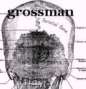
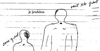
JAMES CURTIS JENKINS: the other laboratory technologist who worked with the autopsy team on JFK, Jenkins was at that time in a Ph.D. program in pathology. ( High Treason II , p. 226) The HSCA's Jim Kelly and Andy Purdy reported that Jenkins "said he saw a head wound in the "...middle temporal region back to the occipital." (HSCA interview with Curtis Jenkins, Jim Kelly and Andy Purdy, 8-29-77. JFK Collection, RG 233, Document #002193, p.4) He told author, David Lifton, "I would say that parietal and occipital section on the right side of the head--it was a large gaping area...It had just been crushed, and kind of blown apart, toward the rear." (Lifton, Best Evidence ", p. 616) When Lifton told Jenkins that photographs showed that the back of the head was essentially intact, except for a small bullet entry wound at the top, he responded, "That's not possible, That is totally--you know, there's no possible way. Okay? It's not possible." ( Best Evidence , p. 617) Jenkins told Livingstone, "Everything from just above the right ear back was fragmented...there was (an absence of scalp and bone) along the midline just above the occipital area....this (wound) would not have been low enough to have gotten into the cerebellum." ( High Treason II , p. 228). Jenkins' views, whether as given by the HSCA, Livingstone, or Lifton, are noteworthy by their consistency, and as Jenkins was in a Ph.D. pathology program, his anatomic specificity is of value.
|
||||
| EDWARD REED: one of two X-ray technicians who worked with Jerrol F. Custer taking X-rays told author David
Lifton that he formed an opinion the night of the autopsy that JFK had been shot from the front because the skull
wound was "more posterior than anterior". (Lifton, David, Best Evidence, p. 619) |
||||
|
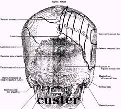
JERROL CUSTER: Q: Now, on the drawing that you have made, and with the bones as theyre identified here, none of the principal
part of the missing wound goes into the occipital bone; is that correct?
|
||||
| JAN GAIL RUDNICKI: Dr. Boswell's lab assistant on the night of the autopsy, Rudnicki was interviewed by
HSCA's Mark Flanagan on 5/2/78. Flanagan reported Rudnicki said, the "back-right quadrant of the head was
missing." (HSCA rec # 180- 10105-10397, agency file number # 014461, p.2.)He told author Harrison Livingston,
"...from the ear back, the scalp was either gone or definitely destroyed in that area.....it would look more
like it was an exit than an entrance." When asked if there was any scalp left in the right rear of the head
behind the ear, Rudnicki said, "That was gone."( High Treason II , p. 207) Rudnicki's account to the
HSCA squares with Livingstone's. |
||||
| JAMES E. METZLER: was a hospital corpsman, third class who helped transport the body from the casket to
the autopsy table in the morgue. Author David Lifton reported, "It was also his impression, from the way the
wound was located toward the back of the head, that President Kennedy must have been shot in the head from the
front." ( Best Evidence, p. 633-634) "Metzler recalled a wound situated in the "right side of the head behind the right ear, extending down to the center of the back of the skull." Metzler mentioned that pieces of brain extended outward from the defect, which measured at least four inches long." (HSCA telephone interview Apr 21, 1978) |
||||
| DAVID P. OSBORNE, M.: a military physician present at the autopsy who was not questioned by the Warren Commission
or the HSCA, he wrote researcher Joanne Braun on 4-5-90 that, "...a second (bullet) hit in the occipital region
of the posterior skull which blew off the posterior top of his skull and impacted and disintegrated against the
interior surface of the frontal bone just above the level of the eyes. I know this for a fact because I was the
one who worked on his head, removing his brain and closed the skull so that he could have had an open casket funeral
if so desired." (Copy of letter furnished to Gary Aguilar by Joanne Braun.) ... "It appeared that the bullet hit low in the occiput of the back of the head and entered the skull there and then traversed a portion of the brain and then hit the inside of the top of the skull toward the rear also and blew a good portion of that part of his skull right out I asked: "Did you actually see the little entry at the bottom of the back of the head?" "Couldn't see the entry," be replied. "That tissue was all pretty much blown away. . . ." I gathered that Osborne based his conclusion that the bullet struck from the rear on an interpretation of where it hit the inside of the skull on the way out I asked him how the bullet could enter from the rear and blow out the rear of the head. He said: ". . . he had to be leaning forward, and the bullet had to hit him in the lower-right behind, you know, that little lump in the back of your head there Osborne was referring to the external occipital protuberance, where Hurnes said there was an entrance wound. Again, I asked Admiral Osborne if he saw that wound. He replied: "Well, the pieces were all blown apart, so it didn't make One tiny little hole in the bone-no. . . . it blew that portion of the skull into Several pieces. L1FTON: I see. So you didn't actually see an entry wound, per se, but it's inferred that it was somewhere towards the bottom of that big hole-or something like that? OSBORNE: It had to be. Otherwise it couldn't have hit the inside of the skull where it did." (lifton 656 657 pbk) |
||||
|
JOHN EBERSOLE, MD: was Assistant Chief of Radiology and head of the Radiology Division at Bethesda, and was the radiologist who evaluated the X-rays in close cooperation with the autopsists on the night of the autopsy. He was not called to testify before the Warren Commission. However he was called to testify by the HSCA on March 11, 1978. Ebersole's deposition was not published by the HSCA causing it to be sealed for 50 years under congressional rules. (Due to pressure, however, the transcript of his interview was released in October, 1993.) A brief wire service account appeared regarding his appearance before the HSCA claiming that he agreed with the Warren Commissions' conclusions. However, in an interview with reporter Gil Dulaney published two days before his HSCA appearance Ebersole said of the head wound, "When the body was removed from the casket there was a very obvious horrible gaping wound to the back of the head (BE:543).", and "The front of the body, except for a very slight bruise above the right eye on the forehead, was absolutely intact. It was the back of the head that was blown off." (BE:546)
|
||||
|
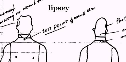
RICHARD A. LIPSEY: an aide to General Wehle who was Commanding General of the military District of Washington, U. S. Army, he was present at JFK's autopsy. In an interview with the HSCA's Andy Purdy and Mark Flanagan on 1-18-78, he claimed that the autopsists "were 'absolutely, unequivocally' convinced that he (JFK) had been shot three times...there were three separate wounds and three separate bullets.". Lipsey gave a confusing account of JFK's head wound. He "identified the entrance in the lower head as being just inside the hairline", but claimed that there was "no real entrance in the rear of the head...one bullet blasted away an entire portion (entrance and exit)..." (sic). Purdy also reported that Lipsey felt that "one bullet entered the back of the head and exited resulting in part of the face and head being blown away" (HSCA, JFK Collection, RG 233) Lipsey completed an autopsy face sheet diagram that depicted an area of the right lateral skull missing, anterior and posterior to the ear, where he had written "same area blown away as wound". In addition, there was a wound low in the skull, presumably of entrance, that was the source of the throat exit wound, which he labeled bullet #2. Finally there was a wound on the back, labeled #3 but the bullet could not be found in the body Lipsey claimed.
|
||||
| PHILIP C. WEHLE: then Commanding officer of the military District of Washington, D. C., he described
the head wound to the HSCA's Andy Purdy on 8-19-77 He did not describe it to the Warren Commission. A copy of memo
on Purdy's interview with Wehle was only released in 1993. Purdy reported that Wehle said he was an observer during
the later stages of the autopsy. "(Wehle) noticed a slight bruise over the right temple of the President but
did not see any significant damage to any other part of the head. He noted that the wound was in the back of the
head so he would not see it because the President was lying face up; he also said he did not see any damage to
the top of the head, but said the President had a lot of hair which could have hidden that...."
(HSCA record # 10010042, agency file # 002086, p. 2) |
||||
| CAPTAIN JOHN STOVER: then Commanding Officer of the National Naval Medical School, he gave no description
of the skull wound to the Warren Commission. (The Pathology Department was under the jurisdiction of the school.)
The HSCA's Mark Flanagan reported that he interviewed him and, "Stover observed...a wound on the top of the
head...".(HSCA Document received from C. Cunningham, 10-22-92) |
||||
| CHESTER H. BOYERS: Boyers "was stationed at Bethesda naval hospital and was the chief Petty Officer
in charge of the Pathology Department in November 1963." (HSCA Telephone contact--Mark Flanagan, 4/25/78,
rec #? 13614) Flanagan reported, "In regard to the wounds Boyers recalls an entrance wound in the rear of the head to the right of the external occipital protuberance which exited along the top, right side of the head towards the rear and just above the right eyebrow." (HSCA Telephone contact--Mark Flanagan, 4/25/78, rec #? 13614, p. 2) |
||||
FLOYD ALBERT REIBE : a medical photographer at Bethesda "JFK: An Unsolved Murder", KRON, 11/18/88
(repeated in "JFK: The Case for Conspiracy" video 1993 [see still photo on p. 88 of Groden's "TKOAP"])---"a
big gaping hole in the back of the head. It was like somebody put a piece of dynamite in a tin can and lit it off.
There was nothing there."; Strongly disagreed with the autopsy photos: "The two pictures you showed
me are not what I saw that night." Interviewer: "What did it look like?" "It had a big hole
in it. This whole area was gone
It's being phonied someplace. It's make-believe.";
|
The Headwound As Seen At Parkland Hospital